
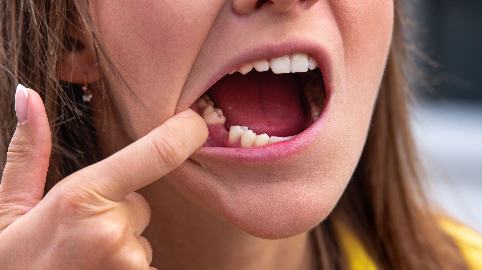

When an extraction becomes complex—deep impaction, multiple teeth, medical risks, or a procedure expected to last hours—you may need general anesthesia to ensure safety, comfort, and surgical efficiency.
General anesthesia is typically reserved for the most difficult cases where local or sedation won’t control pain, anxiety, or airway and where the surgeon needs an immobile, well-controlled field. A practice like Inland Institute Oral & Maxillofacial Surgery is equipped to evaluate and manage these situations safely.
You’ll learn what makes an extraction “complex,” how different impaction types change the plan, how clinicians evaluate risks beforehand, and the anesthesia options that match those risks.
The article will also walk through safety steps, what to expect during recovery, and how long-term follow-up protects your oral health.
Table of Contents
ToggleCriteria for Identifying Complex Tooth Extractions
Complex extractions arise when anatomy, tooth position, or patient health increases technical difficulty, operative time, or complication risk.
You should assess bone density, root morphology, proximity to vital structures, and patient medical status to decide on referral, special techniques, or general anesthesia.
Anatomical Factors
Evaluate root morphology first: dilacerated, fused, hypercementosed, or unusually long roots raise fracture risk and may require sectioning or bone removal.
Roots that are ankylosed to alveolar bone significantly increase force needed and likelihood of adjacent bone loss.
Assess surrounding bone quality and thickness.
Dense cortical bone or limited buccal plate access can limit tooth elevation and increase operative time.
Note thin alveolar plates or sinus proximity; these alter flap design and raise sinus communication risk.
Identify nearby vital structures: inferior alveolar nerve, mental foramen, maxillary sinus, and nasal floor.
If roots extend near or into these structures, plan imaging-guided approaches, possible coronectomy, or involve an oral surgeon.
Tooth Positioning Challenges
Impaction depth and angulation dictate complexity.
Fully impacted third molars that are horizontal, distoangular, or located below the mandibular canal require bone removal and sectioning more often than vertical, superficially impacted teeth.
Eruption status matters.
Partially erupted teeth with limited soft-tissue access or teeth covered by bone need more extensive soft tissue and osseous management.
Supernumerary, ectopic, or transposed teeth often sit in atypical locations that complicate exposure and closure.
Consider root proximity to adjacent roots or restorative work.
Teeth with divergent roots interlocked with neighboring tooth roots or with posts and crowns nearby demand delicate extraction to avoid damaging adjacent teeth or restorations.
Patient-Specific Risk Considerations
Medical comorbidities influence your anesthesia and surgical plan.
Bleeding disorders, anticoagulant use, uncontrolled diabetes, and severe cardiopulmonary disease increase perioperative risk and may necessitate hospital-based extraction under general anesthesia.
Behavioral and cognitive factors affect cooperation.
Severe dental anxiety, autism spectrum disorder, or inability to tolerate local anesthesia can make in-office management unsafe or impractical, prompting consideration of general anesthesia.
Assess past surgical history and medication effects.
Prior radiation to the head and neck, bisphosphonate or antiresorptive therapy, and immunosuppression raise risk of osteonecrosis and infection.
Plan conservative techniques, specialist referral, or modified perioperative protocols accordingly.
Types of Impactions and Their Clinical Implications
Impacted teeth vary by position and tissue coverage, and each type influences extraction difficulty, anesthesia choice, and oral health risks.
Knowing whether a tooth is covered by soft tissue, bone, or partially erupted helps you and your surgeon plan imaging, surgical approach, and postoperative care.
Soft Tissue vs. Bony Impaction
A soft-tissue impaction occurs when the crown penetrates the alveolar bone but remains covered by gum tissue.
You may see swelling, localized pain, or a flap of gingiva (operculum) that traps food and bacteria.
Soft-tissue cases often allow simpler access and can be removed under local anesthesia if other factors (age, anxiety, medical history) are favorable.
Bony impaction means the tooth remains at least partly encased in alveolar bone.
These require bone removal and tooth sectioning more often than soft-tissue cases.
You should expect longer operative time, greater postoperative swelling, and higher likelihood of general anesthesia or deeper sedation when multiple teeth or complex angulations are involved.
Partial Eruption Concerns
Partial eruption leaves the crown exposed but not fully aligned with the occlusal plane.
That exposed margin creates a nidus for bacterial colonization and recurrent pericoronitis.
You may experience intermittent pain, difficulty opening, and foul taste or drainage when infection develops.
Partially erupted teeth complicate flap design and wound closure during extraction.
They also increase the chance of needing sectioning or apical curettage.
Discuss with your surgeon whether preoperative antibiotics, chlorhexidine rinses, or scheduling under sedation will improve outcomes in your specific case.
Associated Oral Health Risks
Impacted teeth can damage adjacent structures: they push against neighboring roots, cause resorption, or destabilize periodontal support.
You should be alert for root resorption of the second molar, angled impactions that alter bite forces, and cyst formation radiographically around the crown (dentigerous cysts).
Impaction also raises caries risk on both the impacted tooth and the distal surface of the preceding tooth.
Chronic inflammation increases the chance of localized bone loss and may complicate later restorative or orthodontic plans.
Your treatment choice—monitoring, surgical exposure, relocation, or extraction—depends on these documented risks and your overall dental plan.
Assessment and Preoperative Evaluation
You will assess anatomy, systemic risk, and likely intraoperative challenges to decide if general anesthesia is required.
Focused imaging, thorough medical history, and predicting specific complications guide your choice of anesthetic modality and surgical plan.
Diagnostic Imaging Techniques
Use panoramic radiography (OPG) for an initial overview of impaction depth, angulation, and developing adjacent tooth relationships.
Panoramic images quickly show third molar position, vertical vs. horizontal impaction, and proximity to the inferior alveolar canal.
Order cone-beam CT (CBCT) when panoramic views suggest close neurovascular relationships, irregular root morphology, or sinus involvement.
CBCT provides three-dimensional views of root curvature, cortical bone thickness, and true distance to the mandibular canal or maxillary sinus.
Obtain periapical radiographs for localized detail when roots, periodontal status, or periapical pathology could alter the surgical approach.
Document imaging findings in the chart and annotate measurements that influence anesthesia and surgical time estimates.
Medical History Review
Screen for cardiovascular, pulmonary, hepatic, renal, and bleeding disorders because these conditions affect anesthetic choice and perioperative risk.
Ask specifically about hypertension, ischemic heart disease, COPD/asthma, chronic kidney disease, cirrhosis, and coagulopathies.
Review current medications and supplements, including anticoagulants, antiplatelet agents, bisphosphonates, SSRIs, and herbal products like ginkgo or garlic.
Determine whether to continue, adjust, or bridge therapies in consultation with the patient’s physician.
Assess airway history and physical exam elements: Mallampati score, neck mobility, mouth opening (interincisal distance), and presence of TMJ disease or obstructive sleep apnea.
Document allergies, previous anesthetic complications, and social factors such as ability to comply with postoperative restrictions.
Predicting Complications
Estimate the risk of nerve injury by correlating root morphology and CBCT proximity to the inferior alveolar or lingual nerve.
Note curved or divergent roots, intimate canal contact, and radiographic darkening as higher-risk features.
Anticipate bleeding and infection risks by combining medical history with local findings: anticoagulant use, liver disease, uncontrolled diabetes, and existing periodontal or periapical infection increase complication likelihood.
Plan hemostatic measures and perioperative antibiotics accordingly.
Project operative difficulty and duration from angulation, depth, bone density, and root shape.
If predicted surgical time, patient anxiety, or expected blood loss exceed safe thresholds for local techniques, favor general anesthesia and prepare appropriate airway management and perioperative monitoring.
Anesthesia Options for Dental Extractions
You will encounter three main approaches to manage pain and anxiety for extractions: targeted numbing that keeps you awake, sedation that reduces awareness while maintaining basic reflexes, and deep anesthesia that renders you unconscious.
Each option varies by monitoring needs, recovery time, and suitability for complex or impacted teeth.
Local Anesthesia Overview
Local anesthesia numbs the extraction site using injections such as lidocaine or articaine.
You remain fully awake and responsive; the goal is to eliminate pain while preserving protective airway reflexes.
Local blocks and infiltrations target specific nerves (e.g., inferior alveolar nerve block for lower molars).
Onset is typically rapid, and duration ranges from 1 to 4 hours depending on the agent and any vasoconstrictor used.
You should expect normal sensations to return gradually and follow instructions on biting and eating until numbness resolves.
Local anesthesia is usually sufficient for simple extractions and many surgical removals when anxiety is manageable.
Conscious Sedation Approaches
Conscious sedation reduces anxiety and awareness but allows you to respond to verbal commands.
Options include nitrous oxide (laughing gas), oral sedatives (e.g., benzodiazepines), and IV sedation (e.g., midazolam, propofol at sedative doses).
Nitrous oxide provides rapid onset and quick recovery; you breathe it through a mask and remain cooperative.
Oral sedatives are easy to administer but have variable absorption and longer recovery; arrange a ride home.
IV sedation produces predictable, titratable effects and is preferred for more complex extractions when you need deeper relaxation without full anesthesia.
Continuous monitoring of oxygenation, heart rate, and blood pressure is required for IV sedation.
General Anesthesia Indications
General anesthesia renders you fully unconscious and requires airway management and advanced monitoring.
It suits cases where multiple teeth are removed, extensive bone removal is needed, or when you have severe anxiety, cognitive impairment, or medical conditions that prevent cooperation.
Your surgical team evaluates medical history, airway anatomy, and bleeding or cardiac risks before recommending general anesthesia.
Procedures under general anesthesia occur in a hospital or accredited surgical center with an anesthesiologist or CRNA present.
Recovery from general anesthesia takes longer and includes close post-anesthesia monitoring for breathing, nausea, and pain control.
Plan for transportation, fasting beforehand, and clear pre-op instructions from the provider.
Determining the Necessity of General Anesthesia
You should weigh patient behavior, the technical demands of the procedure, and any medical risks that rule out lighter options.
These three areas determine whether general anesthesia (GA) provides safer, more effective care than local anesthesia with sedation.
Patient Anxiety and Cooperation Issues
If you have severe dental phobia, past traumatic experiences, or panic disorder that prevents cooperation, GA often becomes the safest option.
High anxiety can make local anesthesia unreliable because movement or sudden reactions increase the risk of soft-tissue injury, incomplete extraction, or airway compromise.
Assess behavioral history and previous responses to sedation.
Consider failed conscious sedation, inability to tolerate injections or prolonged mouth opening, and age-related factors—young children or patients with developmental disabilities commonly need GA.
Document psychological diagnoses, current medications (especially anxiolytics or antidepressants), and any protective behavioral techniques already tried.
Use a brief checklist during evaluation:
- Prior sedation outcomes (success/failure)
- Observable cooperation during exam
- Communication ability and comprehension
- Comorbid psychiatric conditions and medications
Surgical Complexity Factors
You should choose GA when the extraction requires maneuvers that are prolonged, extensive, or carry high risk of complications.
Examples include full-mouth extractions, multiple impacted third molars, deeply impacted canines, or cases requiring alveoloplasty or bone removal exceeding routine levels.
Complexity markers:
- Number of teeth and expected operative time (>2–3 hours often favors GA)
- Depth and position of impaction on imaging
- Need for osteotomy, sectioning, or inferior alveolar nerve manipulation
- Anticipated blood loss or airway management challenges
If imaging shows roots near the mandibular canal or maxillary sinus, or if you expect extended surgical steps, GA improves immobility and surgical field control.
Coordinate with an anesthesiologist and surgical team when multiple complexity markers are present.
Medical Contraindications to Alternatives
You should rule out moderate/deep sedation or local anesthesia when medical conditions elevate risk.
Significant respiratory disease, severe obstructive sleep apnea, uncontrolled gastroesophageal reflux, or severe cardiovascular instability can make lighter sedation unsafe.
Specific contraindications include:
- Inability to maintain a patent airway while sedated
- Severe obesity with apnea-related desaturation
- Recent myocardial infarction or unstable angina
- Coagulopathy that complicates emergency airway or bleeding control
Also review medications that interact with sedatives (e.g., high-dose benzodiazepines, certain opioids).
If you find any of the above, document the contraindication and discuss GA in an appropriate surgical setting—hospital or ASC—with perioperative monitoring and airway management resources.
Safety Considerations and Risk Management
You should expect continuous physiologic monitoring, clear protocols for common complications, and specific postoperative instructions to reduce bleeding, infection, and pain.
Staffing, airway planning, and medication choices directly affect your safety and recovery.
Monitoring During Anesthesia
You will have continuous pulse oximetry, noninvasive blood pressure measurements every 3–5 minutes, and ECG monitoring throughout the procedure.
Capnography must be used when you receive general anesthesia or deep sedation to confirm ventilation and detect hypoventilation early.
Anesthesia providers will maintain a visible airway plan that may include endotracheal intubation or supraglottic airway devices depending on tooth position and expected duration.
Temperature monitoring and urine output assessment are added for longer cases or if significant blood loss is anticipated.
Medication records list induction agents, opioids, muscle relaxants, and local anesthetic doses.
Communicate allergies, home medications, and recent fasting times so providers can anticipate interactions and aspiration risk.
Common Adverse Events
The most frequent immediate risks are nausea, vomiting, sore throat, and transient hypoxia. You may also experience bleeding from the extraction site, which requires local pressure and sometimes sutures or hemostatic agents.
Less common but serious complications include airway obstruction, aspiration, cardiovascular events (arrhythmia, hypotension), and adverse drug reactions. If multiple impacted teeth are removed or the surgery lasts over two hours, infection and delayed bleeding risk increases.
Risk factors that raise complication likelihood include obesity, obstructive sleep apnea, poorly controlled cardiovascular or pulmonary disease, anticoagulant use, and limited mouth opening. Tell your team about these issues so they can adjust monitoring intensity and perioperative medications.
Postoperative Recovery Guidelines
You should remain in a monitored recovery area until you meet discharge criteria: stable oxygen saturation >95% on room air, controlled pain, minimal bleeding, and an alert mental state. Plan for a responsible adult to escort you home and stay with you for 24 hours if general anesthesia was used.
Follow clear oral care steps: bite on gauze for 30–60 minutes to control bleeding. Avoid spitting or using straws for 48 hours, and resume gentle rinsing with saline after 24 hours.
Take prescribed antibiotics and analgesics exactly as directed. Stop or adjust anticoagulants only if instructed by the prescribing clinician.
Seek immediate care for persistent bleeding not controlled with pressure, fever >38°C (100.4°F), increasing facial swelling, uncontrolled pain, difficulty breathing, or new neurologic symptoms. Keep your postoperative instructions and emergency contact numbers accessible.
Recovery and Postoperative Care After General Anesthesia
You will spend a short period in recovery while staff monitor your breathing, heart rate, and responsiveness. Expect grogginess and mild nausea; these usually pass within a few hours.
Have a responsible adult drive you home and stay with you for the first 24 hours. Avoid operating machinery, signing legal documents, or making major decisions during this time.
Follow the written post-op instructions your surgeon provides. These typically include pain control, antibiotic guidance, and wound care to reduce infection risk.
Pain and swelling often peak around 48–72 hours after surgery. Use prescribed analgesics, apply cold packs for the first 24–48 hours, and switch to warm compresses afterward if advised.
Oral hygiene precautions protect the surgical site. Rinse gently with saline or an antiseptic mouthwash as instructed.
Avoid vigorous spitting or using a straw for 7–10 days to prevent dry socket.
Watch for warning signs: uncontrolled bleeding, fever over 38°C (100.4°F), severe or worsening pain, or numbness that does not improve. Contact your surgeon or emergency services promptly if any of these occur.
Quick-reference checklist
- Arrange a caregiver for 24 hours
- Take medications as directed
- Keep head elevated and rest
- Follow diet recommendations: soft foods, avoid hot/spicy items
- Attend scheduled follow-up appointment
Long-Term Outcomes and Follow-Up Strategies
You should expect most patients to regain normal function and comfort within weeks to months after complex extractions under general anesthesia. Healing timelines vary with age, medical history, and the complexity of impaction.
Monitor for common late complications such as dry socket, sensory disturbances, and delayed infection. These issues typically present within the first few weeks but can persist.
Document symptoms and compare with baseline sensory and functional assessments. Schedule structured follow-up visits at 7–10 days, 1 month, 3 months, and 6 months postoperatively to track healing and intervene early.
Use a combination of clinical exam and patient-reported outcome measures, such as pain and chewing ability. Employ targeted imaging when symptoms persist.
Provide clear written and verbal aftercare instructions covering oral hygiene and activity limits. Include signs that warrant urgent review.
Emphasize adherence to analgesic and antibiotic regimens when prescribed. Discuss the expected course for numbness or paresthesia.
Consider long-term rehabilitation for patients with persistent masticatory dysfunction or altered occlusion. Referral options include prosthodontics, physiotherapy, or neurosensory re-education depending on the deficit.
Key monitoring checklist (use at each visit):
- Wound healing and soft-tissue integrity
- Pain score and analgesic usage
- Sensory testing of inferior alveolar/lingual nerves
- Occlusion and masticatory performance
- Need for imaging or specialist referral
Document all findings and patient communications to guide future care decisions and to support informed consent for any additional interventions.