
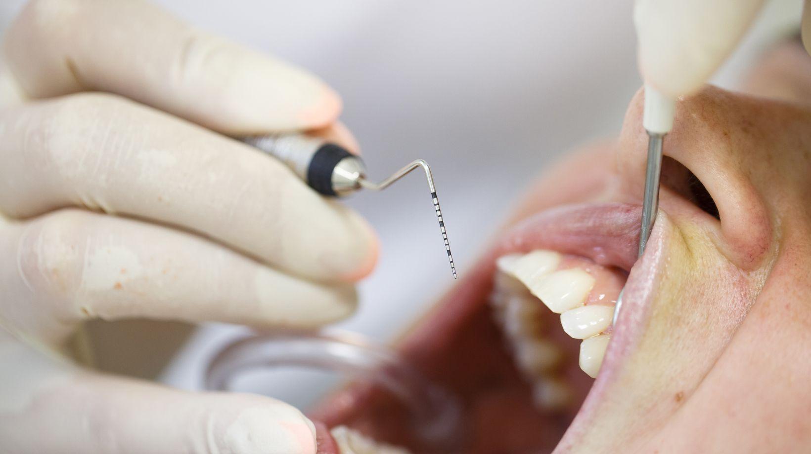

Many patients in Winterville cannot move forward with dental implants because unhealthy gums and infected bone must be treated first to create a stable foundation for the implant to last.
If you have active gum disease or bone loss, your chance of implant failure, infection, or complications rises sharply unless a periodontist treats those issues before implant placement. Practices like Beacon Dental can help coordinate this kind of periodontal evaluation before moving forward.
You’ll learn how periodontal disease undermines implants and what evaluations and treatments specialists use to fix the problem.
This article walks through the biological reasons, practical steps, and local factors that determine whether implants can safely follow periodontal therapy.
Table of Contents
ToggleUnderstanding Periodontal Health in Dental Implant Candidates
Your gum and bone condition determine whether implants can move forward.
Addressing infection, tissue loss, and inflammation first protects implant stability and long-term success.
Risk Factors for Gum Disease in Winterville Residents
Local lifestyle and health patterns influence your risk.
Tobacco use, including cigarettes and vaping, raises your chance of periodontitis and slows healing after any oral surgery.
Diabetes—common in many adults—increases your susceptibility to infection and impairs gum-tissue repair; poorly controlled blood sugar is a major red flag for implant planning.
Oral hygiene habits matter: inconsistent brushing, infrequent flossing, and skipping professional cleanings let plaque mature into destructive biofilm.
Medications that reduce saliva, certain immunosuppressants, and a history of heavy alcohol use also increase risk.
Previous periodontal disease or significant bone loss around teeth directly raises the likelihood of complications after implants, so your clinician will screen for these before approving implant placement.
Clinical Signs of Poor Periodontal Health
Look for bleeding on brushing or probing; it signals active inflammation.
Receding gums and longer-looking teeth indicate attachment loss and expose root surfaces vulnerable to decay and sensitivity.
Increased tooth mobility or shifting teeth suggests supporting bone loss; this often requires intervention before implants.
Persistent bad breath and pus around the gumline point to infection that must be eradicated first.
Deep periodontal pockets (typically measured at 5 mm or more) on probing exams show disease severity and predict higher implant risk if untreated.
Your dentist will document pocket depths, radiographic bone levels, bleeding on probing, and mobility to make a treatment decision.
Early Diagnosis and Its Importance
Early detection preserves bone and simplifies treatment.
When you catch periodontal disease at the gingivitis or mild periodontitis stage, non-surgical therapy—scaling and root planing plus improved home care—often stabilizes tissues without major surgery.
Timely diagnosis prevents progression to advanced bone loss that would require grafting or make implants unfeasible.
Your clinician will use full-mouth probing, digital radiographs, and medical history to stage disease and tailor a plan.
Identifying and controlling systemic contributors, like diabetes or smoking, improves healing and raises the chance of implant success once periodontal health is restored.
Biological Foundations of Implant Success
Healthy soft tissue, adequate bone volume, and controlled inflammation form the biological basis for predictable implant placement.
These elements determine whether the implant will integrate, remain stable under function, and resist infection over time.
Role of Healthy Gums in Osseointegration
Your gums create the first line of defense around an implant and form the soft-tissue seal that limits bacterial penetration.
Firm, keratinized gingiva adapts tightly to the implant abutment and reduces micro-movement and bacterial colonization at the implant–soft tissue interface.
If you have thin, non-keratinized tissue, you face higher risks of recession and exposure of implant components.
That exposure increases contamination risk and may compromise the bone–implant contact needed for osseointegration.
Clinically, this often requires soft-tissue grafting or site augmentation before placing the implant.
Key points to monitor:
- Width of keratinized tissue — ideally ≥2 mm
- Tissue thickness — thicker biotype favors stability
- Mucosal attachment level — stable margins reduce pocketing
How Inflammation Impacts Surgical Outcomes
Active periodontal inflammation elevates local cytokines and proteases that degrade connective tissue and bone.
If you have unresolved periodontitis, the same pathogens and inflammatory mediators that damaged your teeth can colonize implant surfaces and drive peri-implant mucositis or peri-implantitis.
Inflammation also impairs healing by reducing blood flow and interfering with clot stability at the surgical site.
This increases the chance of impaired osseointegration, early implant failure, and the need for additional surgery.
For predictable outcomes, clinicians often require infection control and a period of maintenance until inflammation markers and clinical bleeding scores normalize.
Practical measures before implant surgery:
- Eliminate active pockets and bleeding on probing
- Achieve controlled plaque scores
- Verify stable periodontal probing depths over at least one maintenance cycle
Tissue Regeneration Requirements
Successful implants need sufficient vertical and horizontal bone to achieve primary stability and long-term support.
You may require ridge augmentation, bone grafting, or sinus elevation when residual bone height, width, or density is inadequate for the planned implant dimensions.
Soft-tissue volume matters for aesthetics and barrier function.
Where gingival contours are deficient, connective tissue grafts or collagen matrices restore thickness and reduce recession risk.
Regeneration strategies depend on defect type:
- Horizontal defects: guided bone regeneration with particulate graft + membrane
- Vertical defects: staged grafting or distraction osteogenesis for predictable gain
- Sinus pneumatization: lateral or crestal sinus lift with grafting materials
Your clinician will evaluate clinical probing, CBCT imaging, and occlusal demands to design the appropriate regenerative sequence before implant placement.
Consequences of Untreated Periodontal Issues Prior to Implant Placement
Untreated gum infection and active bone loss change how implants behave in your mouth and raise the chance of needing more complex care.
The following subsections explain specific risks to implant survival, healing, and the jawbone you must know before proceeding.
Increased Risk of Implant Failure
If you have active periodontitis, bacteria and inflammation persist around remaining teeth and can colonize the implant surface after placement.
This raises the risk of peri-implantitis, which can progress faster than natural tooth periodontitis and lead to implant mobility or loss.
Smokers and patients with poorly controlled diabetes face even higher failure rates when periodontal disease is untreated.
Your dentist will assess pocket depths, bleeding on probing, and radiographic bone levels because these factors predict lower implant survival if not stabilized first.
Addressing periodontal infection prior to implant surgery reduces the probability of early and late implant failure and often improves long-term success of both single implants and full-arch restorations.
Post-Surgical Complications
Active gum disease increases the chance of poor wound healing, persistent infection, and need for additional procedures after implant placement.
You may experience prolonged swelling, pain, or drainage that requires antibiotics, surgical cleaning, or implant removal.
Contamination of the surgical site with periodontal pathogens makes grafts and membranes less likely to integrate, increasing the odds of graft failure and repeat surgeries.
Your clinician may delay implant placement until you complete periodontal therapy and demonstrate stable clinical parameters to minimize these complications.
Follow-up maintenance becomes more demanding; untreated disease often necessitates more frequent professional cleanings and monitoring to prevent recurrence around implants.
Effects on Bone Structure
Periodontitis causes progressive bone loss around natural teeth; the same destructive process can affect bone around implants if microorganisms and inflammation remain.
Reduced bone volume complicates implant planning: you may need bone grafts, sinus lifts, or shorter/wider implants to achieve primary stability.
Severely resorbed ridges can change the prosthetic design, increasing cost and treatment time.
Even after grafting, ongoing inflammation reduces graft success rates and can cause further bone loss, compromising esthetics and function.
Your dentist will evaluate bone height, width, and quality with radiographs or CBCT scans and typically require disease stabilization before attempting augmentation or implant placement.
Comprehensive Periodontal Evaluation Procedures
This evaluation identifies infection, bone support, pocket depths, and systemic risks that affect implant success.
Expect detailed probing, targeted imaging, and a review of health factors that influence healing and long-term outcomes.
Clinical Assessment Techniques
You will receive a full periodontal charting that records probing depths, bleeding on probing, recession, and tooth mobility.
- Probing depths: measured at six points per tooth to map pockets and detect attachment loss.
- Bleeding on probing: indicates active inflammation and infection risk.
- Recession and furcation involvement: document root exposure and multi-root defects that may need grafting or altered implant planning.
- Tooth mobility and occlusal assessment: identify teeth that compromise bone or bite forces.
Clinicians also inspect plaque, calculus, and soft-tissue phenotype (thin vs. thick biotype).
These findings determine whether non-surgical therapy, surgery, or staged implant timing is required.
Digital Imaging and Diagnostic Tools
You will get targeted radiographs and, when indicated, a CBCT scan to evaluate bone volume and anatomy.
- Periapical and bitewing X-rays: show bone height, periodontal bone loss patterns, and periapical pathology.
- CBCT (cone beam CT): measures ridge width, identifies anatomic limits (sinus, nerve), and maps bone density for implant positioning.
- Intraoral scans and digital models: help plan prosthetic outcomes and surgical guides.
- Photographs: document soft-tissue contours and esthetic concerns.
Combining these tools gives precise measurements for grafting needs, implant diameter/length selection, and whether immediate placement is feasible.
Patient Medical and Dental History Review
You must provide a complete medical and dental history because systemic conditions change treatment priority and timing.
- Medications: bisphosphonates, anticoagulants, immunosuppressants, and steroids affect healing and infection risk.
- Systemic diseases: uncontrolled diabetes, osteoporosis, and smoking status increase implant failure and periodontal progression.
- Previous periodontal therapy and response: guides likelihood of success with conservative measures.
- Oral hygiene habits and compliance: determine whether non-surgical therapy can stabilize disease before surgery.
The clinician integrates history with clinical and imaging findings to decide if you need periodontal treatment first, what type, and how long to wait before placing implants.
Treatment Strategies to Prepare for Dental Implants
You need controlled gum inflammation, adequate bone volume, and stable oral hygiene before implants.
Treatments range from non-surgical cleaning and antibiotics to surgical pocket reduction and bone grafting, chosen based on disease severity and your health.
Non-Surgical Periodontal Therapies
Non-surgical care focuses on removing bacterial deposits and reducing pocket depths to stop further bone loss.
Your dentist or periodontist will perform scaling and root planing (deep cleaning) to remove plaque and calculus below the gumline; this is often done over one or more visits under local anesthesia.
Adjuncts may include localized antibiotics placed into pockets, antimicrobial rinses, and a short course of systemic antibiotics if infection is widespread.
You’ll receive personalized oral hygiene coaching—brushing technique, interdental cleaning, and frequency—to maintain results.
After treatment, your provider schedules more frequent maintenance cleanings (every 3–4 months) to monitor stability.
If pockets and inflammation resolve and bone loss is minimal, implant planning can proceed.
Surgical Approaches for Advanced Cases
Surgery becomes necessary when non-surgical therapy cannot reduce deep pockets or when bone loss threatens implant stability.
Common procedures include flap surgery to access and debride roots, guided tissue regeneration to rebuild periodontal support, and soft-tissue grafts to thicken or reposition gum tissue for better implant margins.
If you lack sufficient jawbone for implant anchorage, your clinician may recommend bone grafting (autograft, allograft, or xenograft) or ridge augmentation.
Sinus lifts are used for upper molar areas with inadequate vertical height.
Surgical plans often include a healing timeline: 3–6 months for graft integration before implant placement is attempted.
Your systemic health, smoking status, and diabetes control affect healing and the choice of surgical technique.
Collaboration Between Specialists in Implant Cases
Specialists coordinate diagnosis, disease control, surgical timing, and restorative planning so you get a stable foundation for implants and a predictable final result.
Periodontists and General Dentists Working Together
You and your general dentist rely on a periodontist to manage active gum disease and prepare tissues for implants.
The periodontist performs deep cleanings, pocket reduction, or regenerative procedures when bone loss or inflammation threatens implant success.
Your general dentist evaluates occlusion, prosthetic needs, and long-term esthetic goals; that input guides the periodontist’s surgical approach and timing.
Teams commonly share radiographs, periodontal charts, and 3D scans to plan bone grafting, sinus lifts, or staged extractions.
Expect written treatment sequences and clear division of tasks: the periodontist treats disease and optimizes bone and soft tissue, while the dentist designs and places the final restoration when healing is complete.
Communication With Patients on Timeline and Expectations
You should receive a realistic timeline that separates disease control, surgical healing, and prosthetic phases.
Typical steps include 4–12 weeks for periodontal therapy response, 3–6 months for bone graft maturation, and additional healing after implant placement.
The team explains possible delays caused by persistent inflammation, smoking, uncontrolled diabetes, or inadequate bone volume.
Ask for specific milestones and signs of treatment progress, such as reduced pocket depths, stable radiographic bone levels, and soft-tissue contouring before restoration.
Insist on written care instructions and follow-up schedules so you know when to resume normal activities and when the restorative dentist will begin shaping the final crown.
Ongoing Maintenance Following Periodontal Therapy and Implants
You must commit to daily plaque control and a tailored recall schedule to protect both natural teeth and implants.
Professional cleanings, clinical exams, and targeted home-care techniques form the backbone of long-term success.
Long-Term Oral Hygiene Recommendations
You should brush twice daily with a soft-bristle brush and a low-abrasive fluoride toothpaste, paying special attention to the gumline and implant restorations.
Use an interdental brush sized to fit the spaces around your implants and natural teeth; floss daily where contacts exist, and consider floss designed for implants if you have prosthetic components.
Add an antimicrobial rinse (chlorhexidine short-term or alcohol-free Listerine as advised) for 1–2 weeks after surgical or acute inflammatory episodes.
If you smoke, enroll in a cessation program—smoking markedly increases implant and periodontal failure risk.
Bring any new or worsening bleeding, swelling, or looseness to your clinician immediately.
Regular Follow-Up and Monitoring Protocols
Expect professional maintenance visits every 3 months initially.
Your clinician may extend intervals to 3–6 months based on stability and risk factors.
Each visit should include plaque scoring, probing around teeth and implants using a non-abrasive probe, peri-implant radiographs when indicated, and removal of hard and soft deposits with instruments safe for implant surfaces.
Your provider will document pocket depths, bleeding on probing, and mucosal recession to detect recurrence early.
If signs of peri-implant mucositis or peri-implantitis appear, plan for more frequent care and targeted therapy to prevent implant loss.
Factors Unique to Winterville NC Affecting Periodontal Care
Local population characteristics and service availability shape whether you need periodontal treatment before implants.
Income, access to specialists, and common health conditions in the area directly influence diagnosis, timing, and treatment plans.
Local Demographics and Oral Health Trends
Winterville serves a mix of families, older adults, and workers who commute to Greenville and nearby towns.
If you are older or have diabetes — both common in the region — you face higher risk for periodontitis that must be controlled before implant work.
Insurance coverage and income levels in your community affect how often you receive preventive care.
When routine cleanings and early treatment are missed, gum disease advances and requires more intensive periodontal therapy before implants are safe.
Racial and ethnic health disparities reported in nearby North Carolina counties can reflect local variations in periodontal disease prevalence.
If you’re part of an underserved group, expect more careful screening and possible pre-implant interventions to reduce infection risk.
Accessibility of Periodontal and Implant Services
Specialists are concentrated in Greenville and larger centers, so you may need referrals or travel for periodontal assessment and surgery.
If you live in Winterville, plan for additional appointments and coordination between your general dentist and a periodontist.
Availability of non-surgical options like scaling and root planing versus surgical grafting influences timing.
You might receive conservative therapy first; if bone or soft tissue loss is significant, clinicians will delay implant placement until periodontal stability and sufficient bone volume exist.
Local clinics’ appointment wait times, acceptance of Medicaid or private insurance, and patient education resources determine how quickly you progress to implants.
Ask your provider about treatment sequencing, expected timelines, and financial options so you know whether periodontal care will precede implant placement.
Resources for Patients Seeking Periodontal Treatment
You can start by contacting local periodontists in Winterville, NC to discuss diagnosis and treatment timing for implants. Periodontists specialize in gum disease and implant-related care, so they can outline whether non-surgical therapy, surgery, or maintenance is needed first.
Ask your dentist or periodontist about financing and payment plans. Many practices offer in-house plans, third-party financing, or sliding-scale options; verify terms and monthly costs before committing.
Use office support tools to stay connected between visits. Clinics often provide nurse or assistant helplines, answering services, and patient portals for messaging, appointment scheduling, and care instructions.
Look for evidence-based programs that emphasize maintenance and prevention. Regular professional cleanings and personalized oral hygiene coaching can improve outcomes.
Find reputable directories and professional groups to compare providers. Resources include national and local listings and the American Academy of Periodontology for credential verification.
Prepare questions before appointments to make the most of consultations. Ask about staging and grading of periodontitis, expected timelines to implant readiness, and follow-up care requirements.
Keep documentation of prior dental records and imaging handy. Sharing X-rays and notes from previous dentists can streamline diagnosis and your treatment plan.